Interact with the Crabs in Your Computer: New GigaDB Imaging Widgets
 Interactive, downloadable and 3D printable scans of newly discovered hermit crabs are now available in GigaDB using MicroCT technology data. In this latest GigaBlog we let you inspect this data too, as well as provide a Q&A with the first author Jannes Landschoff on its potential utility.
A study just published in GigaScience showcases new functionality in our GigaDB database, as well as a novel data driven way of carrying out descriptions of new species, using three-dimensional visual data from hermit crabs using the latest 3D microCT (Micro computed tomography) scanning technology. By making this microCT data publicly available, taxonomists potentially have more time and cost-efficient options for examining and comparing specimens for taxonomic research, as well as providing new opportunities for education and training. To ease access to these data, the authors go beyond just describing the data collection and findings by providing downloadable, interactive files of everything in this study. For interested citizen scientists out there, we are providing new interactive web-based viewers, video clips and 3D printable file formats.
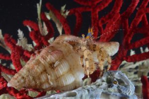 Hermit crabs (Paguroidea), are one of the most diverse superfamilies of sea crustaceans, and are easily recognisable through the way their most-often asymmetrical pleon is concealed and protected usually inside a scavenged sea shell. With over 1,100 species and 120 genera recognised to date, there are many more as yet undiscovered or unidentified species sitting on museum shelves. Being of small size and having very similar body plans and membranous body parts that typically lack identification characteristics, hermit crabs can be extremely difficult to identify and thus remain taxonomically poorly understood. Proper identification requires careful examinations, which depend heavily on the quality of the original species descriptions and illustrations. Much of these procedures have not kept pace with digital advances, and when literature-based descriptions have proven inadequate, the only option has been to contact museums to loan out valuable samples for physical examination.
 New three-dimensional microCT scanning technology provides a new tool in the taxonomic toolkit that can aid with species identification and be consulted by scientists before the physical material has to be sourced and shipped from natural history collections. We’ve recently published best-practice guidelines for this approach as well as examples from brittle-stars, flatworms and snake fangs. Researchers at the University of Cape Town and Stellenbosch University, South Africa, use this approach to scan seven hermit crab samples. This includes three recently described species and one still undescribed species, as well as two scans of rare species, one of which is from a deep-sea habitat at over 500m depth. The most recent species description of Diogenes albimanus has just been published in the journal Zootaxa, where this data was used in the paper and review process.
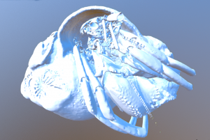
Although the physical specimens used in this study were deposited at the Iziko South African Museum and the Smithsonian National Museum of Natural History, researchers interested in these samples can now inspect them in detail from the comfort of their home. The 3D scans and 3D printer (STL) files are available for download and view from the GigaScience GigaDB repository using widgets that allow embedding in your own website (see the example below and all the others in GigaDB).
Senior Author Anton Du Plessis says of the advantages of X-ray microCT to visualize and analyse these new species: “This study clearly shows the power of the technique and the magnification allows precise identification of surface textures which are often missed in photographs or even in manual drawings.”
GigaScience’s Chris Armit who deployed the new sketchfab viewer in GigaDB said, “The 3D model viewer enables CT and MRI images to be interactively explored. This interface development has huge potential for Digital Biology, and enables morphology and in situ gene expression patterns to be explored easily using a desktop, laptop, or mobile device. We will be extending the visualisation tools to enable exploration of complex image data captured using optical imaging and molecular imaging techniques.”
Following our tradition of author Q&A’s we ask first author Jannes Landschoff a few questions about this work.
How does this microCT approach aid species description, particular in comparison to more traditional ways of describing new species?
MicroCT is still a relatively new technique for taxonomy. For describing new species of hermit crabs, these data were particularly useful to visualize the calcified hard structures using 3D surface-rendered images. A good taxonomic description of a species is highly dependent on quality illustration of the identification characters of the candidate species. Producing drawings of these characters can be difficult, particularly for complex 3D-structures, and drawing artworks are often also subject to the skills of the artist. An ultimate research product though should be reproducible, a condition that requires data transparency. Hence, subjectively-drawn illustrations can violate a basic assumption of scientific experimental design. The peer-reviewing process for taxonomic papers can also be problematic considering that type specimens are usually unavailable, but these are the raw data of the study on which a hypothesis, for example that these specimens belong to a new species, is tested. However, these specimens are too valuable to be shipped, shipping would be too expensive and too slow, and the types can only be examined by one referee at a time. In this process, we used microCT in addition to classical drawings. The scan images hence provided less subjective data, and were particularly suited to provide high-resolution surface imaged of complicated 3D structures that are difficult to draw.
Publishing the dataset also makes the scientific hypothesis of a species being new easier to evaluate, not only in the peer-review process but also in later studies. At the same time these downloadable avatars can act as an insurance policy should the type specimens get damaged or lost.
How useful a tool is microCT in general for the study of hermit crabs?
I think that hermit crabs are a challenging group for CT-scanning, because their pleon is soft, and because they have many fine-scaled soft-tissue ID characters. In my conclusions of my thesis I stated that hermits are maybe the worst possible model organisms within the Decapoda to use CT scanning, yet the scans were valuable. In a nutshell, if hermit crabs can be scanned, other groups like the Brachyura, which are entirely calcified would be very suitable for scanning.
References
du Plessis A, Broeckhoven C, Guelpa A, le Roux SG. Laboratory x-ray micro-computed tomography: a user guideline for biological samples. Gigascience. 2017 Jun 1;6(6):1-11. doi: 10.1093/gigascience/gix027.
Landschoff, J; Du Plessis, A; Griffiths, C, L (2018): A micro X-ray computed tomography dataset of South African hermit crabs (Crustacea: Decapoda: Anomura: Paguroidea). GigaScience. doi:10.1093/gigascience/giy022
Landschoff J, Rahayu DL. A new species of the hermit crab genus Diogenes (Crustacea: Decapoda: Diogenidae) from the coast of KwaZulu-Natal, South Africa. Zootaxa. 2018;4379:268–78.
Landschoff, J; Du Plessis, A; Griffiths, C, L (2018): Supporting data for “A micro X-ray computed tomography dataset of South African hermit crabs (Crustacea: Decapoda: Anomura: Paguroidea)” GigaScience Database. http://dx.doi.org/10.5524/100364