GigaScience at Cell Bio 2020 Virtual

Cell Bio 2020 Virtual – the online ASCB|EMBO Meeting – took place on 2-16 December 2020 and offered many invaluable insights on the structural and evolutionary basis of biological systems. After attending in person in 2019 (see the 2019 write-up here) GigaScience Data Scientist Chris Armit attended the virtual meeting last month and provides one in his series of conference reports on how Cell Biology is enhancing our understanding of the fundamental unit of life.
Archaic Genomes and Human Ancestry
In his keynote talk entitled “Archaic Genomics”, Svante Pääbo (Max Planck Institute of Evolutionary Anthropology, Leipzig, Germany & Okinawa Institute of Science and Technology, Onna-son, Japan) presented a fascinating overview of hominid evolution from a genomics perspective. Svante is one of the key founders of paleogenetics, and his team have developed important techniques for isolating and sequencing DNA sourced from ancient specimens of Homo sapiens and other hominid species. A major finding from Svante’s lab is that admixtures of Neandertal and Denisovan DNA are found in living humans today, and this highlights mating between ancestral humans and these now extinct hominid subspecies in deep prehistory. As Svante explained, as Homo sapiens migrated out of Africa 100,000 years ago, they interbred with Neandertals, and this has left a lasting genetic signature in the genomic DNA of Eurasian populations. In East Eurasian populations we see further interbreeding with Denisovans – named after the Denisova Cave in the Altai mountains of Siberia where this extinct hominid subspecies was first discovered – and Svante further reported that two Denisovan populations contributed to Asian populations. Neandertals and Denisovans were themselves related with a common ancestor 381-473 kyr ago, and this highlights the complexity of hominid ancestry.

Whereas these were fascinating insights into archaic genomes, what I found most surprising was the relevance of these extinct hominid genomes to our understanding of COVID-19 disease in the current pandemic. By correlating clinical severity of COVID-19 outcome with haplotype data, Svante was able to infer that there are multiple alleles on Chromosome 12 that Eurasian populations have inherited from Neandertals that protect against COVID-19 disease. In contrast, there are multiple risk alleles on Chromosome 3 that are also inherited from Neandertals, and that associate with a more severe COVID-19 clinical outcome. As Svante further explained, overall the risk haplotype on Chromosome 3 outweighs the protective haplotype of Chromosome 12, and this suggests that the Neandertal contribution to the human genome is a significant aetiological factor in the progression of COVID-19 disease that associates with a poor outcome (see recent GigaBlogs on predicting COVID-19 outcome and the COVID year in review).
Coronary Artery Development and Disease
In the ‘Growth, Pattern and Form’ symposium, Kristy Red-Horse (Stanford University) delivered a fascinating talk entitled “Using the Cell Biology of Embryogenesis to Inform Tissue Regeneration and Repair in the Heart” that offered invaluable insight into the developing vasculature. In the developing embryo, there are key steps in the formation of organ-specific vascular beds, and these include sprouting angiogenesis, which forms an immature plexus. This is followed by remodelling and pruning of the immature plexus and recruitment of vascular smooth muscle cells.
It had been previously thought that artery formation in vivo required blood flow. However, Kristy’s lab observed arterial markers in the coronary plexus prior to blood flow, and the new model that Kristy is proposing is that venous cells of the developing heart undergo “an early cell fate switch” to create a distinct pre-artery subset, and that these pre-artery cells then migrate and coalesce to form the coronary arteries. Using single-cell RNA seq, Kristy’s lab has shown this convincingly in the mouse whereby expression of the marker gene CX40, which is indicative of pre-artery cells, is observed in the immature vascular plexus prior to blood flow.
So with this in mind, the question then becomes, what is the role of blood flow in the formation of arteries? To investigate this, Kristy’s team looked at the role of a gene called Dach1, which is required for coronary artery development. Dach1 is expressed in all developing coronary endothelial cells, and knockout mice deficient in Dach1 develop smaller coronary arteries, highlighting a critical role in this tissue. Kristy expanded on this through an elegant series of cell culture experiments. In response to laminar flow – which models blood flow in the developing embryo – pre-arterial cells align and elongate in the direction of flow. However, if Dach1 is overexpressed in these cells, there is an enhanced response to laminar flow and the pre-arterial cells migrate against the direction of flow. What is extremely interesting is that Kristy showed that this process is also observable in vivo, and that Dach1 overexpression in endothelial cells “potentiates migration against blood flow in developing arterial blood vessels”.

So why is this important? Coronary artery disease (CAD) is the number one cause of death worldwide, and a major contributing factor to this statistic is the lack of regeneration in the mammalian heart. Using a mouse model of myocardial infarction, Kristy was able to show that overexpression of Dach1 in coronary endothelial cells provides protection against myocardial infarction and decreases mortality. As Kristy explained, “widespread overexpression of Dach1 increases the number of small arteries and improves outcome post-myocardial infarction”. From a translational perspective, this is exciting cell biology research that offers some hope for treating coronary artery disease.
Morphogenesis and Mechanosensation
In the ‘Dynamic Intracellular Organization’ symposium, I was deeply impressed by the presentation by Manuel Théry of the French Alternative Energies and Atomic Energy Commission (CEA, Paris) on “Microtubule Mechano-sensations”. Manuel is a member of the CytoMorpho lab that utilises striking visuals to highlight the role of microtubule self-repair in cells, and to showcase recent experiments that detail mechano-sensitive properties of microtubule networks. Microtubules are polymers of tubulin, and in living cells one can observe that microtubule filaments are constantly growing and shrinking, giving shape to cells and enabling cell movement. Whereas most attention has been focused on the highly dynamic microtubule ends, Manuel was swift to point out that “tubulin dimers can also be exchanged in protofilaments along the microtubule shaft, thus repairing the microtubule and protecting it from disassembly.” As Manuel further explained, “we applied controlled deformations to living cells and found that compressive forces can stabilise microtubules. This process allows the microtubule network to sense and adapt its architecture to the force field cells are submitted to, a process that has long believed to be the apanage of the actin network.” A core concept here is that, in addition to being key structural elements of the cell, microtubules may additionally function like sensory antennae detecting changes in the intracellular and extracellular environment.
The Endosymbiotic Theory Revisited: Endosymbiosis in Action
It is just over 50 years since Lynn Margulis published her seminal cell biology paper “On the origin of mitosing cells”, which proposed a theory of the origin of eukaryotic cells that stated that fundamental organelles, such as mitochondria, photosynthetic plastids, and the basal bodies of flagella “were themselves once free-living (prokaryotic) cells” that were engulfed by a host cell to form a community of endosymbionts that ultimately evolved into organelles. The discovery that mitochondria and chloroplasts have genomes that are distinct from the nucleus, and that these mitogenomes and plastomes are closely related to the genomes of α-proteobacteria and cyanobacteria has lent immense support to the endosymbiotic theory such that it is now widely accepted.

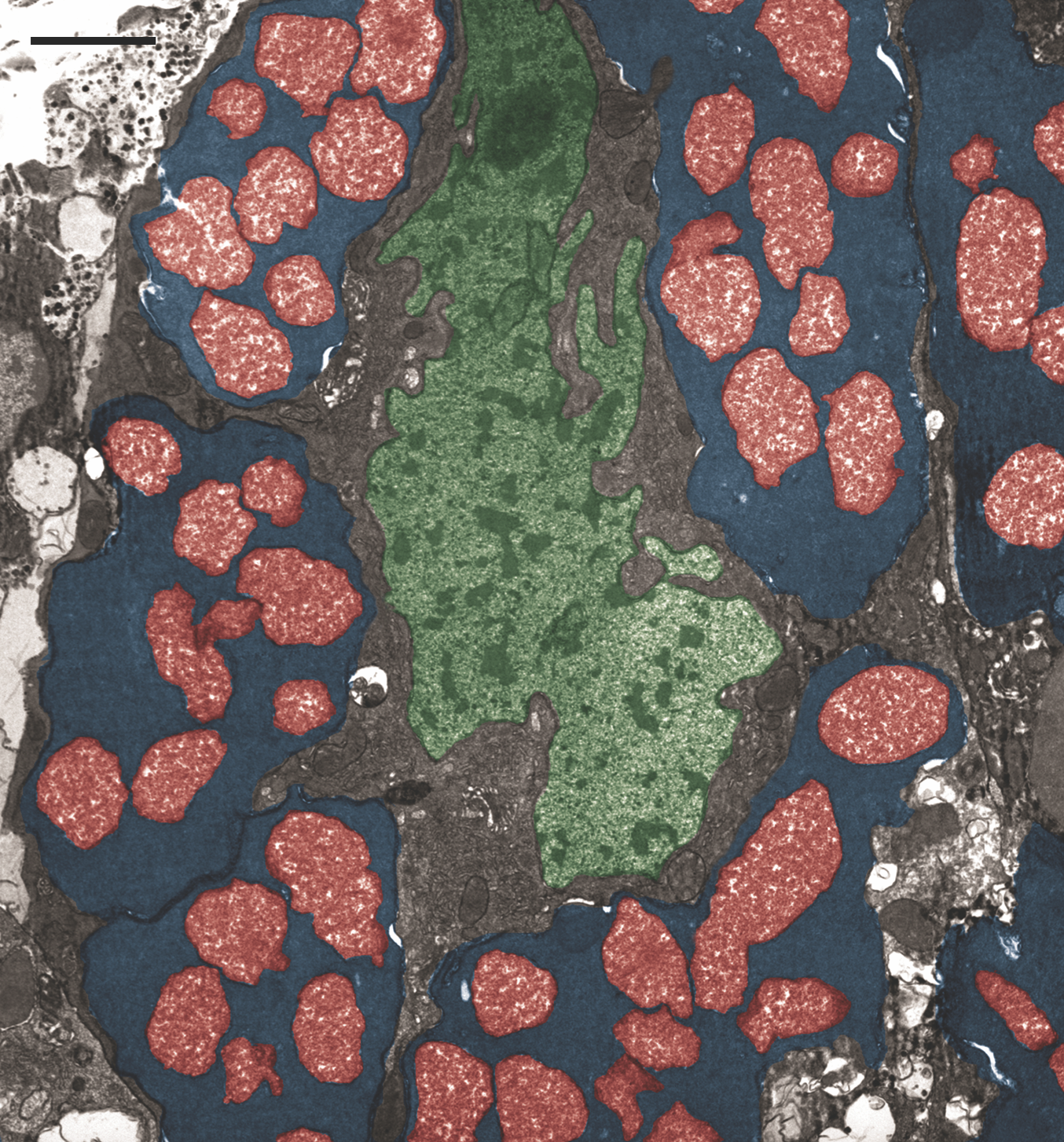
So how could organelles evolve from free-living bacteria? I was intrigued by the talk by John McCutcheon (Arizona State University) entitled “From endosymbionts to organelles: genetic, biochemical, and cell biological integration of bacteria into host cells”. John’s talk was in the subgroup “When Branches of the Tree of Life Meet: Cell Interactions Between Organisms,” and he used the plant sap-eating insect known as the ‘mealybug’ as a model organism for exploring endosymbiosis in action. As John explained, mealybugs obtain all their nutrition from plant sap, but this means that they are lacking essential amino acids in their diet. So how do mealybugs obtain these essential amino acids? The solution is bacterial endosymbionts, which can metabolically generate the missing amino acids that the mealybug cannot make for itself and that it cannot obtain from its very limited diet. These bacterial endosymbionts include species of Tremblaya (Betaproteobacteria) and Moranella (Gammaproteobacteria). What is most surprising is the ‘Russian Doll’ relationship of the endosymbionts, whereby by electron microscopy one can observe that Moranella lives inside Tremblaya, which lives inside the mealybug host cell (see Figure). This “bacterium-in-a-bacterium-in-an-insect cell symbiosis” generates a ‘metabolic patchwork’ that can generate essential amino acids, such as tryptophan. John also highlighted that peptidoglycan synthesis is additionally coordinated in a symbiotic fashion. Peptidoglycan is a component of bacterial cell walls, but is not found in animal cells. However, in the mealybug, the host insect genome is controlling peptidoglycan synthesis for the resident endosymbiotic bacterial populations, and has even acquired some bacterial peptidoglycan (PG)-related genes. These genomes work together to produce a peptidoglycan layer exclusively at the Moranella cell periphery. As John pointed out, this is “a striking parallel to the genetic and biochemical mosaicism found in organelles” and is a fascinating example of endosymbiosis in action.
Mapping Single-Cell Atlases
In his talk entitled “Mapping single-cell atlases across the animal tree of life unravels cell type evolution”, Alexander Tarashansky (Stanford University) highlighted an impressive transcriptome mapping approach that is providing essential detail on the evolution of cell types. Single cell RNA sequencing (scRNAseq) allows near-complete maps of cell types to be generated fairly swiftly. However, as Alexander explained, molecular evolution blurs cell type similarities, and as a consequence using gene expression to map between cell types of distantly related species is very difficult. To address this issue, Alexander and colleagues have developed a Self-Assembling Manifold algorithm, and have utilised this to map cell types across long evolutionary distances. This approach – which Alexander calls SAMap (Self-Assembling Manifold Mapping) – has enabled cell type mapping between fish and frogs. One of the most notable findings of this study was an abundance of ‘paralogue substitutions’ whereby key genes in related cell types in fish and frogs display a more similar expression profile to their paralogue – which is a gene created by a gene duplication event – than to their orthologue – which is derived from a common ancestral gene. This highlights an inherent evolutionary plasticity that needs to be taken into account when transcriptionally profiling cell types in distantly related species.
More ambitiously, Alexander and colleagues have further used SAMap to deliver a pan-species connectivity graph of seven evolutionary distant species – sponge, hydra, schistosoma, flatworm, zebrafish, African-clawed toad (Xenopus), mouse – and this pan-species analysis identified densely connected cell type families. As Alexander explains, by comparing all seven species from sponge to mouse, “we identified interconnected cell type families broadly shared across animals, including contractile and stem cells, along with their respective gene expression programs”. Comparing genetic signatures across these species provides an evolutionary framework that allows us to understand cell type diversity across the Tree of Life (see recent GigaBlog on updates from sequencing the tree of life).
The Curious Relationship of Enhancers and TADs
In “The Genome” symposium, Leonid Mirny (MIT) delivered a thought-provoking talk entitled “Loop Extrusion with Barriers as a Genomic Communication System”. Leonid utilised simulation data to help us understand loop-resolution chromosome conformation capture data, such as that captured by Hi-C. Chromosome conformation capture is a method used to identify the 3D spatial relationship of chromosomes within eukaryotic cells, and it utilises formaldehyde crosslinking of chromatin to capture a snapshot of the spatial relationship between chromosomal loci. The genomic DNA is then digested with a restriction enzyme, and, for Hi-C, the fragments are sequenced. The spatially adjacent cross-linked fragments represent self-interacting genomic regions that are known as topologically-associated domains (TADs), and these can be visualised using a 2D proximity map. TAD boundary regions are often enriched in insulator binding protein CTCF (CCCTC-binding factor), and it is thought that these insulating elements improve the fidelity of enhancer-promoter interactions,
Using simulated data, Leonid helped us to more accurately interpret these 2D proximity maps. By using this approach, Leonid highlighted that chromosomal loop extrusion – which is known to be mediated by cohesin – compacts DNA locally, but this is insufficient to generate TADs, and CTCF insulating barriers are additionally required to generate TADs in these simulations. CTCF insulating barriers, when introduced to the simulated data, robustly generate TADs with narrow-range contacts generating sharper ‘masks’ in the 2D proximity maps. However, even in simulated data these insulated barriers are not absolute, and Leonid invited caution whereby intra-TAD domains are to be interpreted as chromosomal domains where one observes an increased likelihood of locus-to-locus interaction.

So in living systems, what happens when TAD boundaries are removed? In “The Genome” symposium, Wendy Bickmore (MRC Human Genetics Unit, Institute of Genetics and Molecular Medicine, Edinburgh) addressed this very question in her talk entitled “The Role of Spatial Proximity in Genome Regulation.” In collaboration with Bob Hill and Laura Lettice – also based in the MRC Human Genetics Unit – Wendy highlighted the consequences of removing the TAD boundaries that envelop the ZRS enhancer – a long-range enhancer that is solely responsible for Shh expression in the limb – and the Shh promoter. Deletion of CTCF site 1 (ΔCTCF1) – which delineates the TAD boundary 3’ of Shh – resulted in the Shh locus losing interactions with the rest of its own TAD. This was observed using a chromosome conformation assay (5C) and confirmed using FISH. Similar observations were seen for deletion of additional CTCF sites (ΔCTCF2, ΔCTCF3), and it was hypothesised that deletion of these CTCF insulating barriers would have a knock-on effect of mis-regulating Shh gene expression in the limb, perhaps even leading to limb abnormalities similar to what is observed when the ZRS enhancer locus is mutated in the mouse. Surprisingly, deletion of these CTCF sites did not have an impact on gene expression in the limb, nor did it cause any defects in limb development. As Wendy explained, “altered TAD boundaries do not necessarily perturb enhancer function” The reason for this is currently not understood, but this fascinating project highlights a gap in our knowledge as to the curious relationship between enhancers and TADs.
Next year’s ASCB Meeting is scheduled for December 11-15, 2021, in San Diego.